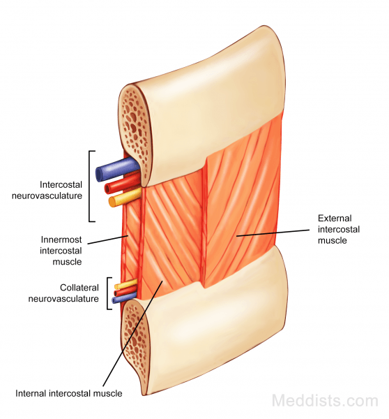
The intercostal neurovasculature passes between the internal and innermost intercostal muscles. It is split into main and collateral neurovasculature.
Description
- The main branches run right under the costal groove (except for first two)
- The collateral branches run at the bottom of the intercostal space
The main branches are arranged in a pattern of VAN — vein, artery, nerve from top to bottom, while the collateral branches are arranged in NAV from top to bottom.
Innervation
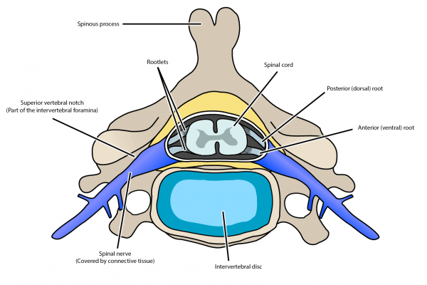
There are 12 pairs of spinal nerves in the thoracic region. All of them leave from the vertebral column through the intervertebral foramina. As soon as they exit, they split into an anterior or ventral ramus and a posterior or dorsal ramus (see Figure 3).