The carpal tunnel is a passage in the wrist formed by the carpal bones and the overlying structures, allowing structures to pass from the forearm to the hand and back.
Carpal tunnel.
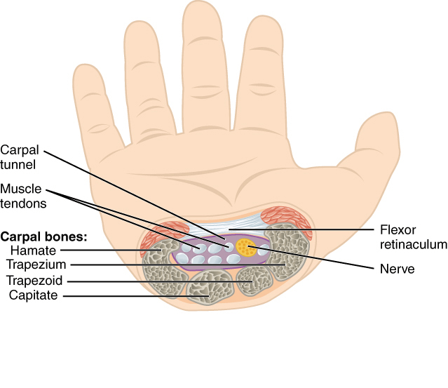
Formed by four carpal bones and the flexor retinaculum, the carpal tunnel forms as a deep arch on the anterior surface of the wrist.
- Medially, it is formed by the pisiform and hook of the hamate bones, while laterally, it is formed by the tubercles of the scaphoid and trapezium bones.
- Both sides of the carpal arch are connected by the flexor retinaculum, a thick layer of connective tissue, forming the tunnel.
- It serves as a pathway for:
- The tendons of the flexor digitorum profundus muscles.
- The tendons of the flexor digitorum superficialis muscles.
- The tendon of the flexor pollicis longus muscle.
- The median nerve.
- These structures are held together by synovial sheaths, one for the flexor digitorum muscles and one for the flexor pollicis longus muscle.
- Other related structures pass into the hand by having a close relation with the carpal tunnel, but without being part of it:
- The tendon of the flexor carpi radialis muscle has its own synovial sheath placed laterally in the flexor retinaculum.
- Anterior to the flexor retinaculum, the ulnar artery and ulnar nerve pass in their own synovial sheath, while the tendon of the